
The way two mpox virus proteins change shape as they bind together acts like a switch that starts the process of viral replication, a study led by researchers at the Blavatnik Institute at Harvard Medical School reveals. This kind of detailed structural information promises to help scientists, armed with a new generation of AI-powered virtual screening tools, target their search for ways to inhibit viral replication, treat mpox and related diseases, and prepare for emerging pathogens. Protein shape shift reveals how the mpox virus begins replicating by Jake Miller, Harvard Medical School edited by Lisa Lock, reviewed by Robert Egan Lisa Lock Scientific Editor Meet our editorial team Behind our editorial process Robert Egan Senior Editor Meet our editorial team Behind our editorial process Editors' notes This article has been reviewed according to Science X's editorial process and policies.
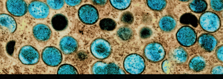
Editors have highlighted the following attributes while ensuring the content's credibility: fact-checked peer-reviewed publication trusted source proofread The GIST Add as preferred source Colorized transmission electron micrograph of mpox virus particles (teal) in an infected cell (brown). "Learning as much as we can about as many different potential threats as we can is the best way to be ready for the pathogens we will face in the future," said study co-senior author Jonathan Abraham, professor of microbiology at HMS. The findings are published in Nature.
Although vaccines are available to help prevent mpox—the disease caused by the mpox virus—and its close relative, smallpox, there is currently no antiviral proven effective against mpox. Mpox is one in a long and ongoing series of emerging and constantly evolving viral threats to humanity, Abraham said. The team made its discovery through a combination of two technologies.
Cryo-electron microscopy showed the structure of the shapeshifting proteins in near-atomic detail at different moments during assembly and activity, while an exquisitely sensitive tool known as optical tweezers showed how the machinery works in real time to unwind the DNA double helix. Mpox has emerged as a global concern The mpox virus was first discovered in animals in 1958, and the first human case was recorded in 1970. The virus is closely related to smallpox, one of the deadliest viruses humanity has ever faced.
Since 2022, an outbreak of mpox has caused more than 100,000 cases in humans and spread to 122 countries, according to the U.S. Centers for Disease Control and Prevention.
Most cases of mpox are relatively mild, but severe cases and complications can cause blindness and even death. "Mpox is caused by a poxvirus, so we know to be afraid of it," Abraham said.
"There's a lot that we need to learn to really be prepared for mpox virus and other viral threats." For example, early in the outbreak that started in 2022, there was hope that the antiviral tecovirimat would be an effective therapy, but it failed to live up to those hopes in a large clinical trial. Treating patients inspires research Abraham, a physician-scientist, also works as an infectious disease doctor at Brigham and Women's Hospital.
The challenges he faces in the clinic help inspire his work in the lab. He's seen firsthand what can happen to people who are sick with viruses for which there is no effective treatment.
While his research is not aimed at finding specific drug targets, Abraham says that broadening and deepening the fundamental understanding of how viruses and other pathogens work is key to accelerating drug discovery. In this case, he was particularly interested in learning how two key proteins work together to allow mpox to replicate—the helicase-primase and the polymerase.
Each member of the duo performs a distinct task. The helicase-primase unzips the virus's DNA double helix and attaches a chemical anchor that allows a new strand of DNA to be built, and the polymerase recruits and organizes the building blocks that assemble into a new strand of DNA.
Together, these pieces are called the replisome. To see what the functioning replisome looked like, the team used cryo-EM data collected at the Harvard Cryo-EM Center for Structural Biology at HMS.
Before the replisome assembles, when the helicase-primase is alone, the primase region of the protein curls up in the center of the helicase, blocking the channel that DNA needs to pass through as it is unwound. But when the functioning replisome assembles, the polymerase binds to the helicase-primase, pulls the primase region out of the channel, leaving the whole unit open and able to function.
Previous work by other researchers had defined the structures of these two proteins, but in biochemical tests, the helicase-primase was practically inert. It couldn't do its job until it worked together with the polymerase.
Medical Disclaimer: This article is for informational purposes only and does not constitute medical advice. Always consult with qualified healthcare professionals for medical decisions and treatment options.
Discover more from ChuckysCarnage
Subscribe to get the latest posts sent to your email.
 ChuckysCarnage