
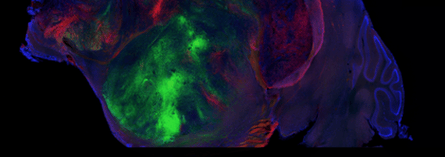
Our brains and their associated diseases are notoriously hard to study. So, researchers developed an animal model with a cortex—the brain’s outer layer—grown from human-derived cells. A side view of a "xenocortical" mouse brain, with the grafted human tissue seen in green and red.
Pașca Lab / Stanford University In a research lab in California, mice that look ordinary on the outside scurry around their cages. But these animals are remarkable on the inside: Roughly half of their brains are made of human nerve cells, or neurons. Scientists transplanted lab-grown human neurons into these rodents, which were genetically engineered to lack parts of their brains.
Specifically, they don’t grow the organ’s outer layer, called the cortex, which is important for things like movement, decision-making and attention. The resulting “xenocortical” mice offer a new way to study brain disorders such as schizophrenia, cerebral palsy and dementia, according to a study published in the journal Nature on September 16. “Our goal has been to make aspects of human brain development and function accessible for investigation, so we can develop therapeutics,” says study co-author Sergiu Pașca, a neuroscientist at Stanford University, to Elie Dolgin at Science News.
Cross-sectional MRI scan of a xenocortical mouse brain showing a map of estimated nerve-fiber pathways. Pașca Lab / Stanford University Human brain diseases are notoriously difficult to study. The organ is extremely complex, containing some 86 billion neurons with trillions of connections between them, and researchers have limited access to living human brain tissue.
That’s why for more than a decade, scientists have been working on tiny replicas of the human brain called organoids. They’re often made from reprogrammed human skin cells.
Previously, Pașca and his colleagues successfully transplanted organoids into rats to study a rare genetic condition called Timothy syndrome. Most people with it also have autism and a heightened risk of epilepsy.
The researchers grew some of the organoids from samples from patients with Timothy syndrome. But they were simply adding the human tissue to the rodents’ existing brain tissue, so the two were competing for space.
The rodent cells’ growth outpaced that of the human cells. This time, the researchers created a way to genetically modify mice so they didn’t develop their cortex or hippocampus, an area critical for learning and memory.
When the animals were 2 days old, they received injections of organoids—each containing roughly 100,000 human-derived cells—in the empty spaces in their brains. “Human cells that are placed in these animals will divide, will grow, and within a few weeks to a few months, they will take most of that space,” Pașca tells Antonio Regalado.
New brain map Earlier this month, researchers released a complete map of an adult male fruit fly’s central nervous system —its brain and nerve cord, the insect equivalent of a human spinal cord. The wiring diagram, which includes 166,700 neurons and more than 300 million connections between them, will help researchers better understand how all brains work, including ours.
By three months after implantation, human tissue had taken over more than 90 percent of the cortex. The cells integrated themselves into the animals’ neural wiring.
Xenocortical mice also performed similarly to control creatures in behavioral tests. Meanwhile, mice that merely had brain chunks missing showed some memory problems and gait differences compared to normal animals.
What’s more, some of the human-derived brain tissue developed into a specialized cell type known as von Economo neurons, which researchers haven’t been able to grow in the lab. These cells are some of the first to die in patients with frontotemporal dementia, a group of neurodegenerative diseases that mainly affect the cortex’s frontal and temporal lobes, areas important for personality, behavior and language.
Pașca now plans to study this form of dementia with xenocortical mice. “We’re trying to see whether mutations that are associated with frontotemporal dementia are making the [von Economo] cells uniquely susceptible to disease,” Pașca tells Carl Zimmer and Carolyn Y.
Medical Disclaimer: This article is for informational purposes only and does not constitute medical advice. Always consult with qualified healthcare professionals for medical decisions and treatment options.
Discover more from ChuckysCarnage
Subscribe to get the latest posts sent to your email.
 ChuckysCarnage